Kidney Stone Relief Pathways: Medical Treatments, Diagnostic Procedures, and Recovery
Introduction
Few medical acute events trigger pain as intense as a migrating renal calculus. The sudden onset of sharp, colicky discomfort in the flank or lower abdomen can instantly halt your routine and leave you searching for rapid medical intervention.This medical guide reviews the full spectrum of kidney stone treatment, detailing the underlying physiology, characteristic symptoms, diagnostic modalities, and interventional procedures available today. You will also find actionable criteria for selecting qualified medical centers and specialists. For educational resources and assistance locating accredited care providers, UrologyHospitals.com offers tools to help patients navigate their urological healthcare decisions.
Understanding Renal Stone Disease
Kidney stones, clinically categorized as renal calculi or nephrolithiasis, are solid accretions formed from mineral salts and organic material within the renal pelvis or calyces. They develop when urine becomes hyper-saturated with insoluble substances, causing solute molecules to precipitate and form microscopic crystalline structures.
Physiology of the Upper Urinary Tract
The upper urinary tract comprises the kidneys and ureters. The kidneys filter metabolic waste products from blood plasma, maintaining electrolyte balance and producing urine. The urine drains into the renal pelvis, flowing downward through narrow, muscular channels—the ureters—into the urinary bladder.
[ Kidneys / Renal Pelvis ]
           │
           ▼
     [ Ureters ]  <-- (Common site of impaction & colic)
           │
           ▼
     [ Bladder ]
           │
           ▼
    [ Urethra ] ──> Output
Stones anchored within the renal parenchyma or calyces may remain asymptomatic. However, when a calculus dislodges and transitions into the ureter, it causes mechanical irritation and luminal obstruction. The resulting pressure buildup within the renal pelvis causes capsule distension and intense smooth muscle spasms, generating severe renal colic.
Varieties of Renal Calculi
Determining the metabolic profile of the calculus is vital for establishing long-term prevention protocols:
- Calcium-Based Calculi (70–80%): Predominantly composed of calcium oxalate or calcium phosphate; strongly linked to metabolic imbalances or high urinary oxalate levels.
- Uric Acid Calculi (5–10%): Develop in persistently acidic urine environments, often associated with gout, metabolic syndrome, or high-purine diets.
- Struvite (Infective) Calculi: Composed of magnesium ammonium phosphate; formed by urease-producing bacterial infections. These can expand rapidly into staghorn structures filling the renal collecting system.
- Cystine Calculi (< 1%): Stem from cystinuria, a hereditary metabolic disorder disrupting renal tubular transport of dibasic amino acids.
Recognized Symptoms and Clinical Warning Signs
While tiny micro-calculi can pass unnoticed through the urinary tract, larger or impacted stones produce characteristic clinical signs:
- Severe Flank and Back Pain: Intense, intermittent discomfort (colic) localized to the costovertebral angle, fluctuating in severity as the ureter contracts.
- Referred Abdominal & Groin Discomfort: As the calculus migrates downward through the ureter, pain radiates toward the ipsilateral lower quadrant, inguinal region, or external genitalia.
- Lower Urinary Tract Symptoms (LUTS): Irritative storage symptoms, including urinary frequency, dysuria (burning during voiding), and urgent voiding sensations when the stone approaches the ureterovesical junction.
- Gross or Microscopic Hematuria: Pink, red, or tea-colored urine resulting from mucosal abrasion along the ureteral lining.
- Nausea and Emesis: Shared autonomic nerve pathways between the kidneys and gastrointestinal tract frequently induce metabolic upset and vomiting.
Emergency Warning: The combination of acute urinary obstruction and systemic infection is a urological emergency. If severe pain is accompanied by high fever, rigors, confusion, or complete anuria (inability to pass urine), seek immediate emergency department care.
Underlying Etiology and Risk Factors
Calculi formation typically involves multiple overlapping metabolic, environmental, and anatomical factors:
- Inadequate Hydration: Insufficient fluid intake lowers total urine volume, raising solute concentration and lowering the solubility threshold of salts.
- Dietary Imbalances: High sodium intake elevates urinary calcium excretion. Excessive consumption of animal proteins increases uric acid while reducing urinary citrate (a natural stone inhibitor).
- Systemic Health Conditions: Primary hyperparathyroidism, distal renal tubular acidosis, Crohn’s disease, and chronic diarrhea alter systemic pH and electrolyte absorption.
- Familial Inheritance: Genetic predispositions affecting intestinal absorption or renal reabsorption of calcium, oxalate, or uric acid.
- Anatomical Anomalies: Structural variations such as ureteropelvic junction (UPJ) obstruction, horseshoe kidney, or medullary sponge kidney promote urinary stasis.
Diagnostic Workup and Imaging
A targeted diagnostic evaluation is essential to confirm stone burden, identify anatomical obstruction, and rule out concurrent pyelonephritis.
Standard Diagnostic Protocols:
- Clinical Assessment: Evaluation of symptom timeline, pain distribution, medical history, and dietary patterns.
- Reagent Strip & Microscopic Urinalysis: Tests for micro-hematuria, urinary pH, nitrites, leukocyte esterase, and mineral crystals.
- Serum Chemistry Profile: Measures serum creatinine, blood urea nitrogen (BUN), electrolytes, calcium, and uric acid levels to evaluate kidney function and systemic metabolism.
- Non-Contrast Computed Tomography (NCCT): The gold-standard diagnostic tool for acute flank pain. It identifies stone location, size, density (measured in Hounsfield units), and distance to the ureterovesical junction with over 95% sensitivity.
- Renal Ultrasonography: A radiation-free alternative that detects hydronephrosis (kidney swelling) and renal calculi, ideal for pediatric or pregnant patients.
Evidence-Based Kidney Stone Treatment Modalities
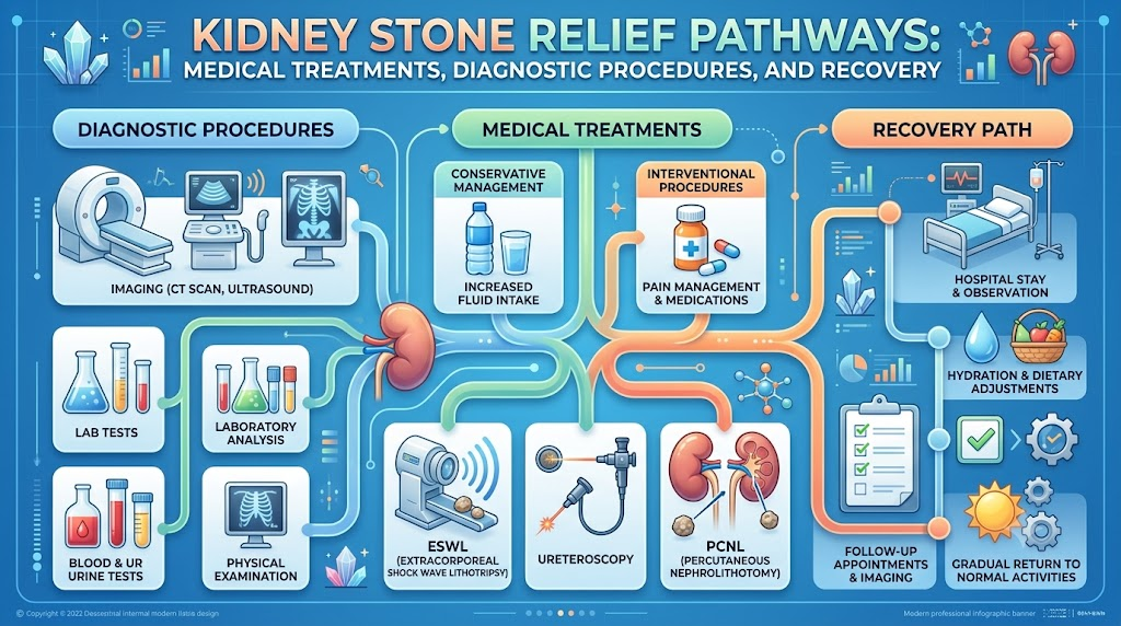
The selected kidney stone treatment pathway is driven by stone size, anatomical position, composition, degree of obstruction, and patient health status.
               [ Diagnostic Imaging (NCCT) ]
                             │
         ┌───────────────────┴───────────────────┐
         ▼                                       ▼
  [ Calculus < 5-7 mm ]                   [ Calculus > 7 mm ]
  Uncomplicated Passage                   Obstructive / Symptomatic
         │                                       │
  ├─ Hyper-hydration                      ├─ Extracorporeal Shock Wave
  ├─ Analgesic Therapy                    │  Lithotripsy (ESWL)
  └─ Alpha-Blocker (MET)                  ├─ Ureteroscopy (URS) + Laser
                                          └─ Percutaneous Nephrolithotomy (PCNL)
1. Conservative & Medical Expulsive Therapy (MET)
For small, non-obstructive calculi (typically under 5–7 mm) without concurrent infection:
- Targeted Hydration: Consuming sufficient fluids to achieve a minimum daily urine output of 2.5 liters helps transport the stone through the urinary tract.
- Analgesia: Non-steroidal anti-inflammatory drugs (NSAIDs) are preferred for first-line pain management, reducing ureteral smooth muscle inflammation and capsular tension.
- Medical Expulsive Therapy: Selective alpha-1 receptor antagonists (e.g., tamsulosin) relax the smooth muscle of the distal ureter, increasing clearance rates and shortening passage time.
2. Extracorporeal Shock Wave Lithotripsy (ESWL)
- Technique: Delivers focused acoustic shock waves generated outside the body through the skin to fragment intra-renal or upper ureteral stones into fine gravel.
- Indications: Solitary radiopaque renal or upper ureteral calculi under 15–20 mm in diameter with favorable renal anatomy.
- Clinical Nuances: Performed as an outpatient procedure under conscious sedation. It is less effective for very dense stones (e.g., monohydrate calcium oxalate or cystine) or obese patients where the shock wave path length is excessive.
3. Ureteroscopy (URS) and Retrograde Intrarenal Surgery (RIRS)
- Technique: A flexible or semi-rigid ureteroscope is advanced through the urethra, bladder, and ureter directly to the stone. A Holmium:YAG or Thulium fiber laser then pulverizes the calculus.
- Indications: Mid-to-distal ureteral stones, ESWL-resistant calculi, or lower pole kidney stones under 20 mm.
- Clinical Nuances: Performed under general anesthesia with high stone-free success rates. A temporary ureteral stent may be placed post-operatively to preserve urinary flow while ureteral swelling subsides.
4. Percutaneous Nephrolithotomy (PCNL)
- Technique: A small tract is established through the posterior flank directly into the renal collecting system. A nephroscope is inserted, and ultrasonic or pneumatic lithotripters break up large stone masses before manual extraction.
- Indications: Large renal calculi (> 20 mm), staghorn stones, or complex lower pole collecting system stones.
- Clinical Nuances: Performed under general anesthesia in a hospital setting, requiring short inpatient observation and specialized post-operative care.
Additional Urological Conditions & Interventions
Complex urological care often intersects with other lower tract conditions that require specialized medical or surgical intervention:
- Enlarged Prostate Treatment: Benign Prostatic Hyperplasia (BPH) can cause bladder outlet obstruction, urinary stasis, and secondary bladder stone formation. Options include pharmacological management, transurethral resection (TURP), or laser ablation.
- Prostate Cancer Treatment: Care plans depend on Gleason scores, clinical stage, and PSA kinetics, spanning active surveillance, radical prostatectomy, radiotherapy, and androgen deprivation therapy.
- Bladder Cancer Treatment: Often presenting with painless gross hematuria, bladder urothelial carcinoma is managed via transurethral resection of bladder tumor (TURBT), intravesical instillations (BCG/chemotherapy), or radical cystectomy.
- Urinary Tract Infection Treatment: Involves targeted antimicrobial regimens based on urine culture sensitivity testing, alongside investigation of predisposing anatomical anomalies.
- Robotic Urology Surgery: Employs computer-assisted robotic platforms to provide high 3D visualization and fine articulation during delicate operations, such as partial nephrectomy or radical prostatectomy.
Modality Comparison Matrix
| Treatment Approach | Primary Indication | Key Benefits | Clinical Considerations |
| Medical Expulsive Therapy | Non-obstructive stones < 5–7 mm | Avoids invasive procedures; low cost | Requires time; risk of unpredictable pain episodes |
| Shock Wave Lithotripsy (ESWL) | Uncomplicated renal stones < 15–20 mm | Fully non-invasive; rapid recovery | Reduced efficacy on dense stones; fragment passage can cause colic |
| Ureteroscopy (URS / RIRS) | Mid/distal ureteral stones; intra-renal stones < 20 mm | High stone-free rate regardless of stone density | Requires anesthesia; potential temporary ureteral stent discomfort |
| Percutaneous Nephrolithotomy (PCNL) | Large (> 20 mm) or complex staghorn calculi | High clearance rates for complex stone burdens | Higher invasiveness; requires inpatient admission and tract healing time |
Practical Scenarios
Scenario A: Outpatient Management of a Distal Ureteral Stone
Case Summary: A 38-year-old male presents with acute right-sided flank pain radiating to the groin. NCCT imaging demonstrates a 4 mm calculus in the distal right ureter without significant hydronephrosis or fever.
Management: The attending physician initiates Medical Expulsive Therapy (tamsulosin daily), prescribes oral NSAIDs for pain control, and advises generous fluid intake. The calculus passes spontaneously on day four without secondary complications.
Scenario B: Surgical Intervention for an Obstructive Proximal Stone
Case Summary: A 54-year-old female presents with persistent left flank pain and nausea. Diagnostics reveal a dense 10 mm calculus impacted in the upper left ureter causing moderate hydronephrosis.
Management: Given the stone size and persistent obstruction, conservative passage is unlikely. The urologist performs a flexible ureteroscopy with Thulium fiber laser lithotripsy. The stone is pulverized into dust, and a temporary double-J stent is placed for five days to ensure drainage.
Patient Decision-Making Framework
Navigating care for renal stone disease involves a systematic progression of clinical decisions:
[ Step 1: Document Symptoms & Timing ]
                  │
                  ▼
[ Step 2: Undergo Diagnostic NCCT / Ultrasound ]
                  │
                  ▼
[ Step 3: Consult a Urological Specialist ]
                  │
                  ▼
[ Step 4: Compare Interventional Options (Risk vs. Stone Clearance) ]
                  │
                  ▼
[ Step 5: Select Facility & Surgical Team ]
                  │
                  ▼
[ Step 6: Complete Post-Treatment Metabolic Evaluation & Prevention Plan ]
Selecting Urology Hospitals and Specialists
Achieving optimal outcomes requires evaluating providers and medical centers based on structural and clinical capabilities:
- Board-Certified Urology Specialists: Confirm that attending surgeons possess dedicated endourology training or extensive volume experience with advanced lithotripsy techniques.
- Advanced Diagnostic Facilities: Select facilities equipped with low-dose non-contrast CT scanners and high-resolution ultrasonic imaging.
- Comprehensive Surgical Suites: Ensure availability of advanced holmium/thulium laser technologies, flexible ureteroscopes, and dedicated Robotic Urology Surgery platforms for complex reconstructions.
- Emergency Intervention Capabilities: Verify that the institution provides 24/7 coverage for urgent ureteral stenting or percutaneous nephrostomy placement during obstructive infection crises.
- Local Care Continuity: When searching for a Urologist Near Me, consider both proximity for emergency access and the availability of post-procedure follow-up care.
Patients can use platforms like UrologyHospitals.com to research hospital capabilities and explore directory information for accredited specialists.
Common Clinical Pitfalls to Avoid
- Dismissing Early Warning Signs: Ignoring mild flank discomfort or transient hematuria can allow silent, progressive urinary obstruction to cause irreversible renal parenchymal damage.
- Relying on Unverified Dietary Remedies: Attempting to dissolve calcium-based calculi using unproven home remedies can delay necessary medical evaluation.
- Incomplete Antimicrobial Regimens: Stopping prescribed antibiotic therapy early for stone-associated UTIs fosters antibiotic resistance and risks recurrent pyelonephritis.
- Neglecting Post-Passage Metabolic Evaluation: Failing to collect passed fragments for laboratory stone analysis prevents targeted dietary and pharmacological prevention plans.
- Assuming Invasiveness Correlates with Success: Opting for major surgical procedures when less invasive options (such as URS or ESWL) yield equivalent clearance rates adds unnecessary risk.
Key Takeaways
- Early Evaluation Preserves Renal Function: Timely diagnosis prevents obstructive hydronephrosis and chronic renal damage.
- Stone Metrics Drive Procedure Choice: Size, anatomical position, density (Hounsfield units), and collecting system structure determine whether observation, lithotripsy, or surgery is indicated.
- Hydration Remains Essential: Achieving a daily urine volume over 2.5 liters is the foundational defense against solute crystallization.
- Metabolic Analysis Prevents Recurrence: Quantitative analysis of passed stones combined with a 24-hour urine metabolic screen identifies individual recurrence risks.
- Seek Specialized Medical Centers: Access to modern endourological technology and experienced surgical teams improves clearance rates and minimizes complications.
Frequently Asked Questions
What scope of conditions fall under urological care?
Urologists treat diseases of the male and female urinary tract (kidneys, ureters, bladder, urethra) as well as conditions affecting the male reproductive system and prostate gland.
When is prompt urological consultation necessary for kidney stones?
Consult a urologist if you experience severe flank pain, gross blood in your urine, persistent vomiting, or symptoms that fail to improve with basic oral pain medications.
What imaging technique provides the definitive diagnosis for renal calculi?
Low-dose non-contrast Computed Tomography (NCCT) is the clinical gold standard, offering high sensitivity and specificity to locate, size, and measure the density of all stone types.
What medical interventions help clear kidney stones?
Small stones are managed with hydration, NSAIDs, and alpha-blockers. Larger stones are treated using Extracorporeal Shock Wave Lithotripsy (ESWL), Ureteroscopy (URS) with laser fragmenting, or Percutaneous Nephrolithotomy (PCNL).
How do urologists manage benign prostatic hyperplasia (BPH)?
BPH is managed through lifestyle adjustments, alpha-blockers, 5-alpha reductase inhibitors, or minimally invasive surgical options such as transurethral resection or laser enucleation.
What factors dictate prostate cancer treatment plans?
Treatment depends on clinical stage, Gleason grade, serum PSA levels, life expectancy, and patient preference, ranging from active surveillance to robotic surgery or radiation.
What signs indicate an urgent urological emergency?
Fever, chills, or systemic illness combined with obstructive urinary pain requires emergency intervention to decompress the urinary collecting system and prevent sepsis.
How does robotic technology assist in urological surgery?
Robotic systems afford operating surgeons high-definition 3D visualization, tremor suppression, and articulated instruments that enhance precision during minimally invasive surgical procedures.
What criteria should guide the choice of a urological hospital?
Key considerations include surgeon case volume, access to modern laser endourology suites, round-the-clock emergency support, and comprehensive metabolic diagnostic services.
How can patients find qualified local urology specialists?
Patients can review physician credentials, verify hospital affiliations, check patient-education resources on platforms like UrologyHospitals.com, and consult primary care referral networks.
Conclusion
Renal stone disease remains a painful and potentially serious condition, but modern endourological techniques offer highly effective pathways to stone clearance and long-term relief. From medical expulsive therapy to advanced laser ureteroscopy and percutaneous surgery, treatment can be tailored precisely to the characteristics of your stone and your overall health profile.Achieving optimal outcomes relies on prompt medical evaluation, clear communication with your clinical team, and a commitment to post-treatment metabolic prevention. Educational platforms like UrologyHospitals.com can help you research conditions, understand procedure options, and find qualified specialists and care facilities.
Leave a Reply